
 Online Bill Pay
 Patient Portal
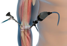
Elbow Arthroscopy
Elbow arthroscopy, also referred to as keyhole or minimally invasive surgery, is a surgical procedure that is performed through tiny incisions to evaluate and treat several elbow conditions.
Biceps Tendon Repair
Biceps tendon repair is a surgical procedure to restore a biceps tendon that has been torn or ruptured by severe trauma or injury.
Endoscopic Cubital Tunnel Release
Endoscopic cubital tunnel release is a minimally invasive surgical procedure to decompress the ulnar nerve for the treatment of cubital tunnel syndrome. Endoscopic refers to the surgery being performed utilizing an endoscope - a thin, flexible fiber-optic tube with a camera, light, magnifying lens, and a port to pass tiny surgical instruments.

Elbow Tendon and Ligament Repair
Ligament reconstruction is considered to treat ligament rupture. Your surgeon will make an incision over the elbow. Care is taken to move muscles, tendons, and nerves out of the way. The donor's tendon is harvested from either the forearm or below the knee.
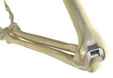
Total Elbow Replacement
Elbow joint replacement, also referred to as total elbow arthroplasty, is an operative procedure to treat the symptoms of arthritis that have not responded to non-surgical treatments. The goal of elbow joint replacement surgery is to eliminate your pain and increase the mobility of your elbow joint.
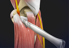
Ulnar Nerve Release
Ulnar nerve release, also known as ulnar nerve decompression, is a surgical procedure to treat a medical condition called ulnar nerve entrapment.
Cubital Tunnel Syndrome (Ulnar Nerve Entrapment)
When the elbow is bent, the ulnar nerve can stretch and catch on the bony bump. When the ulnar nerve is compressed or entrapped, the nerve can tear and become inflamed, leading to cubital tunnel syndrome.
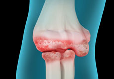
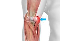
Elbow Arthritis
Although the elbows are not weight-bearing joints, they are considered to be most important for the functioning of the upper limbs. Hence, even minor trauma or disease affecting the elbow may cause pain and limit the movements of the upper limbs.
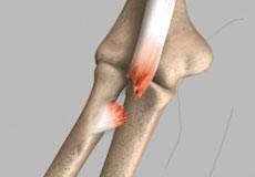
Elbow Fractures
Elbow fractures may occur from trauma, resulting from various reasons: a fall on an outstretched arm, a direct blow to the elbow or an abnormal twist to the joint beyond its functional limit.
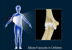
Elbow Fractures in Children
The arm in the human body is made up of three bones that join to form a hinge joint called the elbow. The upper arm bone or humerus connects from the shoulder to the elbow to form the top of the hinge joint.
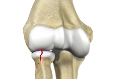
Radial Head Fractures
Radial head fractures are very common and occur in almost 20% of acute elbow injuries. Elbow dislocations are generally associated with radial head fractures. Radial head fractures are more common in women than in men and occur more frequently in the age group of 30 to 40 years.
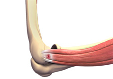
Golfer's Elbow
Golfer’s elbow, also called medial epicondylitis, is a painful condition occurring from repeated muscle contractions in the forearm that leads to inflammation and microtears in the tendons that attach to the medial epicondyle.
Tennis Elbow
Tennis elbow is a common name for the elbow condition lateral epicondylitis. It is an overuse injury that causes inflammation and microtears of the tendons that attach to the lateral epicondyle.
The elbow is a complex joint formed by the articulation of three bones – the humerus, radius, and ulna. The elbow joint helps in bending or straightening of the arm to 180 degrees and lifting or moving objects.
The bones of the elbow are supported by:
- Ligaments and tendons
- Muscles
- Nerves
- Blood vessels
Bones and Joints of the Elbow
The elbow joint is formed at the junction of three bones:
- The humerus (upper arm bone) forms the upper portion of the joint. The lower end of the humerus divides into two bony protrusions known as the medial and lateral epicondyles, which can be felt on either side of the elbow joint.
- The ulna is the larger bone of the forearm located on the inner surface of the joint. It articulates with the humerus.
- The radius is the smaller bone of the forearm situated on the outer surface of the joint. The head of the radius is circular and hollow, which allows movement with the humerus. The articulation between the ulna and radius helps the forearm to rotate.
The elbow consists of three joints, namely:
- The humeroulnar joint is formed between the humerus and ulna and allows flexion and extension of the arm.
- The humeroradial joint is formed between the radius and humerus and allows movements like flexion, extension, supination, and pronation.
- The radioulnar joint is formed between the ulna and radius bones and allows rotation of the lower arm.
Articular cartilage lines the articulating regions of the humerus, radius, and ulna. It is a thin, tough, flexible and slippery surface that acts as a shock absorber and cushion to reduce friction between the bones. The cartilage is lubricated with synovial fluid, which further enables the smooth movement of the bones.
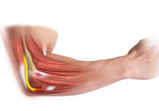
Muscles of the Elbow Joint
There are several muscles extending across the elbow joint that help in various movements. These include the following:
- Biceps brachii: Upper arm muscle, enabling flexion of the arm
- Triceps brachii: Muscle in the back of the upper arm that extends the arm and fixes the elbow during fine movements
- Brachialis: Upper arm muscle beneath the biceps, which flexes the elbow towards the body
- Brachioradialis: Forearm muscle that flexes, straightens and pulls the arm at the elbow
- Pronator teres: Muscle that extends from the humeral head, across the elbow, and towards the ulna, and helps to turn the palm facing backward
- Extensor carpi radialis brevis: Forearm muscle that helps in movement of the hand
- Extensor digitorum: Forearm muscle that helps in movement of the fingers
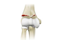
Ligaments and Tendons of the Elbow
The elbow joint is supported by ligaments and tendons, which provide stability to the joint.
Ligaments are a group of firm tissues that connect bones to other bones. The most important ligaments of the elbow joint are the:
- Medial or ulnar collateral ligament: Comprised of triangular bands of tissue on the inner side of the elbow joint
- Lateral or radial collateral ligament: A thin band of tissue on the outer side of the elbow joint
- Annular ligament: Group of fibers that surround the radial head, and hold the ulna and radius tightly in place during movement of the arm
Together, the medial and lateral ligaments are the main source of stability and hold the humerus and ulna tightly in place during movement of the arm.
The ligaments around a joint combine to form a joint capsule that contains synovial fluid.
Any injury to these ligaments can lead to instability of the elbow joint.
Tendons are bands of connective tissue fibers that connect muscle to bone. The various tendons that surround the elbow joint include:
- Biceps tendon: attaches the biceps muscle to the radius, allowing the elbow to bend
- Triceps tendon: attaches the triceps muscle to the ulna, allowing the elbow to straighten
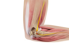
Nerves of the Elbow
The main nerves of the elbow joint are the ulnar, radial and median nerves. These nerves transfer signals from the brain to the muscles that aid in elbow movements. They also carry sensory signals such as touch, pain, and temperature back to the brain.
Any injury or damage to these nerves causes pain, weakness or joint instability.
Blood Vessels Supplying the Elbow
Arteries are blood vessels that carry oxygen-pure blood from the heart to the hand. The main artery of the elbow is the brachial artery that travels across the inside of the elbow and divides into two small branches below the elbow to form the ulnar and the radial artery.